Discover how Intellekt AI's advanced AI solutions revolutionized brain tumor detection from histopathological images, enabling accurate and timely diagnoses for improved patient care.

Key details
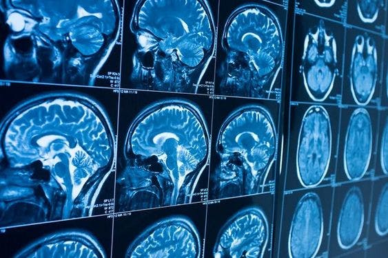
Brain Tumor Detection
Challenge
Accurate and timely detection of brain tumors from histopathological images is crucial for effective treatment. However, manual analysis is time-consuming, subjective, and prone to human errors. There was a need for a reliable and efficient solution to automate the detection process and enhance diagnostic accuracy.
Approach
Intellekt AI implemented a state-of-the-art AI-driven brain tumor detection system. Leveraging deep learning algorithms, the system analyzed histopathological images to identify regions of interest, detect tumor presence, and classify tumor types with high accuracy.
The system was trained on a vast dataset of annotated histopathological images, enabling it to learn complex patterns and distinguish between normal brain tissue and tumor regions. The algorithms were fine-tuned and optimized to achieve exceptional performance and adaptability to different types of brain tumors.
Goal
The goal was to develop an automated brain tumor detection system that could assist pathologists in making accurate diagnoses more efficiently. The aim was to reduce the time required for tumor detection, enable early intervention, and improve patient outcomes.
Outcome
Intellekt AI's AI-powered brain tumor detection system surpassed expectations, delivering remarkable outcomes. The system achieved high sensitivity and specificity in detecting brain tumors from histopathological images, leading to improved diagnostic accuracy and reduced turnaround time.
Brain Tumor Detection
Technologies
Technologies used


Check out other Case Studies

Stock Trading using Deep Reinforcement Learning
Our client was a hedge fund company that uses mathematical formulas to evaluate stocks and their performance. They wanted us to create a more lucrativ...

Recommendation System
Explore how Intellekt AI developed a powerful recommendation system for a finance company, leveraging user behavior patterns to recommend complementar...
Let's discuss a solution for you

Anand Ajmera
will help you with your query
contact@intellektai.comWe are happy to assist you